Loculated Pleural Effusion Cxr / Chest Radiograph Showing A Left Sided Loculated Pleural Effusion Download Scientific Diagram / Differentiation of loculated effusions from solid masses.. A pleural effusion is accumulation of excessive fluid in the pleural space, the potential space that surrounds each lung. Treatment depends on the cause. A loculated pleural effusion can mimic a mass hence is sometimes known as a pleural pseudotumor. Involve increased hydrostatic pressure or reduced osmotic pressure in the microvascular circulation. Determine if it can be tapped.
Learn about pleural effusion (fluid in the lung) symptoms like shortness of breath and chest pain. Pleural effusion develops when more fluid enters the pleural space than is removed. More than one half of these massive pleural effusions are caused by malignancy; In healthy lungs, these membranes ensure that a small amount of liquid is present between the lungs. Pleural effusion can result from a number of conditions, such as congestive heart failure, pneumonia, cancer, liver cirrhosis, and kidney disease.
Pleural Effusion Work Up Youtube from i.ytimg.com A pleural effusion is accumulation of excessive fluid in the pleural space, the potential space that surrounds each lung. Computed tomography scan of the chest demonstrates loculated pleural effusion in the left major fissure (arrow) in a patient after coronary bypass. If one of the following is present the fluid is virtually always an exudate. Other causes are complicated parapneumonic effusion. Learn about different types of pleural effusions, including symptoms, causes learn more from webmd about different types of pleural effusions,including symptoms, causes, and treatments. If none is present the fluid is virtually always a transudate. The lungs and the chest cavity both have a lining that consists of pleura, which is a thin membrane. Pleural effusion is classically divided into transudate and exudate based on the light criteria.
Pleural fluid/serum ldh ratio >0.6.
Terminology pleural effusion is commonly used as. Send aspirated fluid for cytology. Pleural effusions occur as a result of increased fluid formation and/or reduced fluid resorption. Differentiation of loculated effusions from solid masses. Pleural effusion, popularly known as water in the pleura or water in the lung, is the name given to the abnormal accumulation of fluid in the pleura, a thin membrane surrounding the lung. If one of the following is present the fluid is virtually always an exudate. Learn step 2 and shelf essentials in a free 10 min video. Determine if it can be tapped. Pleural effusion is not a disease, but a common manifestation of several different diseases. More than one half of these massive pleural effusions are caused by malignancy; Pleural effusions are abnormal accumulations of fluid within the pleural space. Loculated effusions are mostly due to adhesions driven by pleural inflammation; e intrinsic characteristics of an effusion and its.
Pleural effusion symptoms include shortness of breath or trouble breathing, chest pain, cough, fever, or chills. Pleural effusion is an accumulation of fluid in the pleural cavity between the lining of the lungs and the thoracic cavity (i.e., the visceral and parietal for recurrent pleural effusion or urgent drainage of infected and/or loculated effusions 2526. If none is present the fluid is virtually always a transudate. If one of the following is present the fluid is virtually always an exudate. Learn step 2 and shelf essentials in a free 10 min video.
Epos Trade from epos.myesr.org Pleural effusions may result from pleural, parenchymal, or extrapulmonary disease. Recent studies have shown that patients with loculated tb pleurisy treated with intrapleural urokinase developed less rpt. More than one half of these massive pleural effusions are caused by malignancy; Involve increased hydrostatic pressure or reduced osmotic pressure in the microvascular circulation. Approximately 1 million people develop this abnormality each year in the united states. Pleural effusion, popularly known as water in the pleura or water in the lung, is the name given to the abnormal accumulation of fluid in the pleura, a thin membrane surrounding the lung. Pleural effusion occurs when too much fluid collects in the pleural space (the space between the two layers of the pleura). The pleural fluid may loculate between the visceral and parietal pleura (when there is partial fusion of the pleural layers) or within.
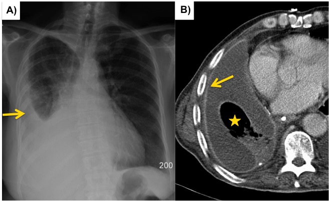
A loculated pleural effusion is the major radiographic hallmark of parapneumonic effusion or empyema (see fig.
Pleural fluid/serum ldh ratio >0.6. Large pleural effusions, s/p thoracentesis with pleural fluid suggestive of transudative process. Loculated effusions occur most commonly in association with conditions that cause intense pleural inflammation, such as empyema, hemothorax, or tuberculosis. Determine if it can be tapped. Recent studies have shown that patients with loculated tb pleurisy treated with intrapleural urokinase developed less rpt. Pleural effusion can be a sign of serious illness. Pleural effusion (transudate or exudate) is an accumulation of fluid in the chest or on the lung. Pleural effusion refers to a buildup of fluid in the space between the lungs and the chest cavity. Pleural fluid/serum protein ratio >0.5. There is a large left pleural effusion obscuring the lower half of the left hemi thorax. If none is present the fluid is virtually always a transudate. Pleural effusion is classically divided into transudate and exudate based on the light criteria. Loculated effusions are mostly due to adhesions driven by pleural inflammation;
Pleural effusion develops when more fluid enters the pleural space than is removed. Determine if it can be tapped. Differentiation of loculated effusions from solid masses. Pleural fluid ldh > two thirds of upper limit for serum ldh. Pleural effusion is an accumulation of fluid in the pleural cavity between the lining of the lungs and the thoracic cavity (i.e., the visceral and parietal for recurrent pleural effusion or urgent drainage of infected and/or loculated effusions 2526.
Role Of Medical Thoracoscopy In The Management Of Multiloculated Empyema Bmc Pulmonary Medicine Full Text from media.springernature.com Homogenous density density in dependent portion upright: Pleural fluid/serum ldh ratio >0.6. Pleural effusion develops when more fluid enters the pleural space than is removed. It detects pleural effusions with higher sensitivity and specificity than cxr, and provides valuable information about the size and depth of the pleural effusion, the echogenicity of the fluid, the presence of septated or loculated fluid, pleural thickening and nodularity, and the presence of any. Loculated pleural effusion on cxr. They may result from a variety of pathological processes which overwhelm the pleura's ability to reabsorb fluid. Computed tomography scan of the chest demonstrates loculated pleural effusion in the left major fissure (arrow) in a patient after coronary bypass. Meaning of pleural effusion medical term.
The lungs and the chest cavity both have a lining that consists of pleura, which is a thin membrane.
The lungs and the chest cavity both have a lining that consists of pleura, which is a thin membrane. Loculated effusions are mostly due to adhesions driven by pleural inflammation; Pleural effusions are a common medical problem with more than 50 recognised causes including disease local to the pleura or underlying lung, systemic conditions, organ dysfunction and drugs. Pleural effusion occurs when too much fluid collects in the pleural space (the space between the two layers of the pleura). Terminology pleural effusion is commonly used as. Pleural effusions can loculate as a result of adhesions. Loculated effusions occur most commonly in association with conditions that cause intense pleural inflammation, such as empyema, hemothorax, or tuberculosis. Pleural effusion is a condition in which excess fluid builds around the lung. Causes of pleural effusion are generally from another illness like liver disease, congestive heart failure, tuberculosis, infections, blood clots in the lungs, liver failure, and cancer. Other causes are complicated parapneumonic effusion. Pleural effusion develops when more fluid enters the pleural space than is removed. Send aspirated fluid for cytology. Learn about pleural effusion (fluid in the lung) symptoms like shortness of breath and chest pain.
Computed tomography scan of the chest demonstrates loculated pleural effusion in the left major fissure (arrow) in a patient after coronary bypass loculated pleural effusion. Pleural fluid/serum protein ratio >0.5.


0 Komentar